Photo contest reveals strange beauty of the microscopic world


Each year, rigorous science and dazzling artistry meet in Nikon’s Small World photomicrography competition.

Started in 1975, the contest celebrates the beauty of images taken through a light microscope. Scientists and hobbyists alike enter, and the winner receives a $3,000 prize. This year, the competition celebrates its 50th anniversary, and it received about 2,100 photo entries from 80 countries.

If sometimes unnerving, the images are always stunning, and this year’s contest is no exception.

1st place

This year’s first place prize was awarded to a groundbreaking image of mouse brain tumor cells, taken by Bruno Cisterna, a faculty member at Augusta University’s Medical College of Georgia. The photo reveals how disruptions in the cell’s cytoskeleton – the structural framework and “highways” known as microtubules – can lead to diseases such as Alzheimer’s and amyotrophic lateral sclerosis (better known as ALS or Lou Gehrig’s disease).

Cisterna’s research was published in May in the Journal of Cell Biology.

Differentiated mouse brain tumor cells (actin, microtubules, and nuclei) (Bruno Cisterna & Eric Vitriol / Medical College of Georgia at Augusta University / Nikon Small World)Differentiated mouse brain tumor cells (actin, microtubules, and nuclei) (Bruno Cisterna & Eric Vitriol / Medical College of Georgia at Augusta University / Nikon Small World)

Bruno Cisterna spent about three months perfecting the staining process to ensure clear visibility of the cells in this image.

2nd place

Second place was awarded to Marcel Clemens, an astronomer turned photographer based in Italy. His image shows an electrical arc between a pin and a wire.

Electrical arc between a pin and a wire (Marcel Clemens (Verona, Italy) / Nikon Small World)Electrical arc between a pin and a wire (Marcel Clemens (Verona, Italy) / Nikon Small World)

Electrical arc between a pin and a wire (Marcel Clemens (Verona, Italy) / Nikon Small World)

3rd place

This image of a cannabis plant leaf, captured by Chris Romaine of Port Townsend, Washington, showcases hairlike plant appendages called trichomes. The bubbles are cannabinoid vesicles — fluid-filled, blisterlike structures.

Leaf of a cannabis plant. The bulbous glands are trichomes. The bubbles inside are cannabinoid vesicles. (Chris Romaine (Port Townsend, Wash.) / Nikon Small World)Leaf of a cannabis plant. The bulbous glands are trichomes. The bubbles inside are cannabinoid vesicles. (Chris Romaine (Port Townsend, Wash.) / Nikon Small World)

Leaf of a cannabis plant. The bulbous glands are trichomes. The bubbles inside are cannabinoid vesicles. (Chris Romaine (Port Townsend, Wash.) / Nikon Small World)

“Sometimes, we overlook the tiny details of the world around us,” said Eric Flem, communications manager at Nikon Instruments. Nikon Small World serves as a reminder to pause, appreciate the power and beauty of the little things, and to cultivate a deeper curiosity to explore and question.”

Scroll through to see other the highlights of this year’s competition:

You say “ladybird,” I say “ladybug”

Ladybug (Angus Rae / Australian National University / Nikon Small World)Ladybug (Angus Rae / Australian National University / Nikon Small World)

Autofluorescence in the face of a little two-spotted ladybird, the British and Canadian name for a ladybug.

Slime mold

Slime mold on a rotten twig with water droplets. (Ferenc Halmos (Hungary) / Nikon Small World)Slime mold on a rotten twig with water droplets. (Ferenc Halmos (Hungary) / Nikon Small World)

Slime mold on a rotten twig with water droplets.

Mosquito larva

Mosquito larva (Anne Patricia Algar (U.K.) / Nikon Small World)Mosquito larva (Anne Patricia Algar (U.K.) / Nikon Small World)

Mosquito larva (Anne Patricia Algar (U.K.) / Nikon Small World)

So many eyes …

Green crab spider		 (Paweł Błachowicz (Poland) / Nikon Small World )Green crab spider		 (Paweł Błachowicz (Poland) / Nikon Small World )

Eyes of a green crab spider.

Crystals

Recrystallized mixture of hydroquinone and myoinositol
 (Marek Miś Photography (Poland)
 / Nikon Small World)Recrystallized mixture of hydroquinone and myoinositol
 (Marek Miś Photography (Poland)
 / Nikon Small World)

Recrystallized mixture of hydroquinone and myoinositol.

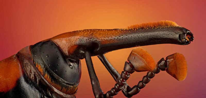
There’s a wasp in there …

An insect egg parasitized by a wasp. (Alison K. Pollack (California) / Nikon Small World)An insect egg parasitized by a wasp. (Alison K. Pollack (California) / Nikon Small World)

An insect egg parasitized by a wasp.

Butterfly wings

Wing scales of a butterfly on a medical syringe needle. (Daniel Knop (Germany) / Nikon Small World)Wing scales of a butterfly on a medical syringe needle. (Daniel Knop (Germany) / Nikon Small World)

Wing scales of a butterfly on a medical syringe needle.

Grimace for the camera!

Transverse section of stem of bracken fern. (David Maitland / St. Andrew's / Nikon Small World)Transverse section of stem of bracken fern. (David Maitland / St. Andrew's / Nikon Small World)

Transverse section of stem of bracken fern.

Reflective eggs

Golden bug eggs on a sage leaf. (Jochen Stern (Germany) / Nikon Small World)Golden bug eggs on a sage leaf. (Jochen Stern (Germany) / Nikon Small World)

Golden bug eggs on a sage leaf.

Water fleas

Two water fleas with embryos, left, and eggs, right. (Marek Miś Photography (Poland)
 / Nikon Small World)Two water fleas with embryos, left, and eggs, right. (Marek Miś Photography (Poland)
 / Nikon Small World)

Two water fleas with embryos, left, and eggs, right.

Feathery antenna

Antenna of a mole crab (Igor Robert Siwanowicz / Howard Hughes Medical Institute / Nikon Small World)Antenna of a mole crab (Igor Robert Siwanowicz / Howard Hughes Medical Institute / Nikon Small World)

Antenna of a mole crab

Hidden world in a grain of sand

Beach sand. (Zhang Chao / Chinese Academy of Sciences / Nikon Small World )Beach sand. (Zhang Chao / Chinese Academy of Sciences / Nikon Small World )

Beach sand.

Slime mold (Part II)

Slime mold (Timothy Boomer / WildMacro.com / Nikon Small World )Slime mold (Timothy Boomer / WildMacro.com / Nikon Small World )

Focus-stacked composite of 337 exposures at 10x lifesize of a slime mold from California’s Eldorado National Forest.

Sea star

Nervous system of a young sea star. (Laurent Formery & Nathaniel Clarke / Stanford University / Nikon Small World)Nervous system of a young sea star. (Laurent Formery & Nathaniel Clarke / Stanford University / Nikon Small World)

Nervous system of a young sea star.

This article was originally published on NBCNews.com


Source link

About The Author

Scroll to Top